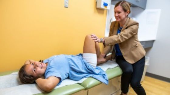
Legg-Calvé-Perthes disease (Perthes) is a painful pediatric hip disorder caused by a blood supply disruption to the femoral head. As the femoral head heals and repairs, it becomes fragile and at risk for
flattening and deformity.
One of the many challenges of treating Perthes is the heterogeneity of the disease, impacting a broad pediatric age group with a spectrum of disease severity. The goal of treatment is to maintain a spherical femoral head and congruent ball-and-socket hip joint during periods of avascularity and subsequent healing. Femoral head deformity correlates with early-onset hip osteoarthritis and the potential premature need for a hip replacement.
The Perthes Research Team at Gillette Children’s, led by Jennifer Laine, MD, and Susan Novotny, PhD, knows that advancing the care of patients who have Perthes requires innovative treatments tailored to the individual case. That’s why the team partnered with researchers Casey Johnson, PhD, Ferenc Tóth, DVM, PhD, and Alexandra Armstrong, DVM, PhD, at the University of Minnesota (UMN) to develop and implement advanced magnetic resonance imaging (MRI) methods to diagnose, assess severity, and evaluate the healing of Perthes. The multidisciplinary team is also collaborating on developing preclinical models of Perthes along with Reza Talaie, MD, at the UMN.
Current Perthes Imaging Limitations
Current clinical management for Perthes predominately relies on conventional radiography (X-ray) for diagnosis and subsequent monitoring. Unfortunately, the radiographic characteristics of Perthes lag behind the physiologic changes by weeks to months. Therefore, an X-ray is not ideal for assessing the extent and location of ischemic injury, nor the rate or extent of revascularization within the femoral head. Despite X-rays being the gold standard tool for managing Perthes, the tool itself cannot comprehensively assess the health of the femoral head when it's most susceptible to permanent deformity.
Another option for imaging the femoral head in Perthes is contrastenhanced MRI (CE-MRI). Unlike X-ray, CE-MRI provides a three-dimensional (3D) assessment of femoral head shape and detects the location and extent of femoral head ischemia and revascularization. However, concerns about the unknown significance of contrast agent deposition in the brain restrict CE-MRI utilization in children with Perthes to their initial diagnosis. The team aims to develop MRI techniques that provide equivalent or improved information about the Perthes hip without using contrast. This would allow more frequent advanced imaging of the hip during the stages of healing, leading to a better understanding of the disease and its repair processes.
Perthes Imaging Innovation
The Gillette and UMN Perthes team has begun developing quantitative, non-CE MRI techniques to monitor the earliest stages of Perthes disease progression and the initial stages of vascular and bone repair.
Quantitative MRI techniques from this study will provide a safe alternative to CE-MRI to enable serial imaging and, therefore, a better understanding of the disease process and healing. This proposed serial imaging will allow researchers to develop more informed clinical treatments and recommendations that reduce the burden of hip osteoarthritis in children with Perthes.
Dr. Novotny says, “Quantitative MRI will provide a more accurate tool for orthopedic surgeons to monitor each child’s hip recovery from ischemic injury. Our families with Perthes are desperately seeking an accurate timeline for when their child will be healed. Quantitative MRI has the potential to quantify the rate and extent of femoral head healing, and therefore inform treatment decisions and evaluate the efficacy of treatments.”
The Future of Perthes Treatments
The Perthes team anticipates that the application of their quantitative MRI techniques, combined with their development of an improved preclinical model of Perthes, will permit a comprehensive characterization of Perthes and its healing. Inducing faster, more efficient healing would improve outcomes. Once both research and clinical innovations are established, the research team can more effectively assess treatments such as non-operative (e.g., non-weight bearing), medical (e.g., biologic agents), and surgical interventions that are tailored to known factors of Perthes.
“In much of medicine, we are working to fine-tune treatment strategies or strengthen prevention efforts,” says Dr. Laine. “In contrast, for Perthes, many of the big questions remain, including why it happens and how best to treat it. This uncertainty is frustrating — and frankly unacceptable — for patients and families dealing with this condition. We can better understand how the hip heals and responds to different treatments by improving our advanced imaging. This will be essential to improve our evidence-based care for patients with Perthes. Our hope is a future for Perthes kids with less pain, better function, and more answers.”
Join Our Partners in Care Community!
Subscribe to Partners in Care Journal, a newsletter for medical professionals.
Subscribe Today Home Page

