What is a craniotomy?
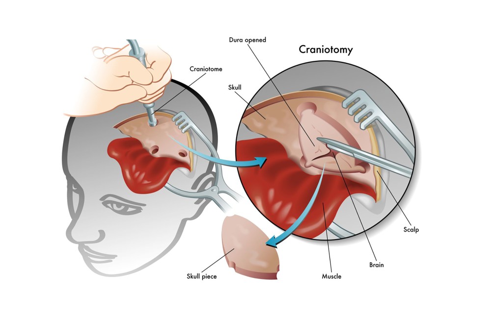
A craniotomy is the surgical removal of part of the bone from the skull to expose the brain. Specialized tools are used to remove the section of bone called the bone flap. The bone flap is temporarily removed, then replaced after the brain surgery is finished.
Some craniotomy procedures may use the guidance of computers and imaging (magnetic resonance imaging [MRI] or computerized tomography [CT] scans) to reach the precise location within the brain that is to be treated. This technique requires the use of a frame placed onto the skull or a frameless system using landmarks on the scalp. When either of these imaging procedures is used along with the craniotomy procedure, it is called a stereotactic craniotomy.
Scans made of the brain during this procedure provide a three-dimensional image of what the brain structure looks like. For example, your provider would be able to see a tumor within the brain with these scans. It is useful in making the distinction between tumor tissue and healthy tissue and reaching the precise location of the abnormal tissue.

Who benefits from a craniotomy?
Because a craniotomy is so useful for viewing the structure of the brain, there are many reasons a doctor may recommend one, including:
- Diagnosing, removing, or treating brain tumors
- Clipping or repairing of an aneurysm
- Removing blood or blood clots from a leaking blood vessel
- Removing an arteriovenous malformation (AVM) or addressing an arteriovenous fistula (AVF)
- Draining a brain abscess, which is an infected pus-filled pocket
- Repairing skull fractures
- Repairing a tear in the membrane lining the brain (dura mater)
- Relieving pressure within the brain (intracranial pressure) by removing damaged or swollen areas of the brain that may be caused by traumatic head injury or stroke
- Treating epilepsy
- Implanting stimulator devices to treat movement disorders or dystonia (a type of movement disorder)
What are the risks of a craniotomy procedure?
As with any surgical procedure, complications may occur from a craniotomy. Brain surgery risk is tied to the specific location in the brain that the operation will affect. For example, if the area of the brain that controls speech is operated on, then speech may be affected. Some more general complications include:
- Infection
- Bleeding
- Blood clots
- Pneumonia (infection of the lungs)
- Unstable blood pressure
- Seizures
- Muscle weakness
- Brain swelling
- Leakage of cerebrospinal fluid (the fluid that surrounds and cushions the brain)
- Risks associated with the use of general anesthesia
The following complications are rare and generally relate to specific locations within the brain, so the risk depends on the individual undergoing the procedure:
- Memory problems
- Speech difficulty
- Paralysis
- Abnormal balance or coordination
- Coma
There may be other risks depending on your specific medical condition. Be sure to discuss any concerns with your doctor before the procedure.
How can I prepare for a craniotomy?
To prepare for a craniotomy procedure, please review the tips to prepare for surgery at Gillette. You can also explore the amenities available at Gillette before your stay.
What can I expect on the day of a craniotomy procedure?
A craniotomy takes place under general anesthesia, which means you’re asleep throughout the procedure. Your neurosurgeon will perform a craniotomy, opening the skull to access the brain. A craniotomy usually takes between 2.5-3 hours.
During the procedure, your surgeon:
- Removes a piece of your skull
- Peels back a section of the dura, the tough membrane that protects the brain
- Uses surgical microscopes to insert special instruments to remove or cut pieces of brain tissue based on the reason for craniotomy
- Replaces the dura
- Uses stitches or staples to secure the skull bone back into place
Every child heals differently, and outcomes depend on the neurologic condition of your child before surgery.
A craniotomy generally requires a hospital stay of 3 to 7 days. You may also go to a rehabilitation unit for several days after your hospital stay. Procedures may vary depending on your condition.
After the craniotomy, your child will spend one to three days in the pediatric intensive care unit (PICU) for close monitoring.
Craniotomy Services at Gillette Children's
If your child experiences a medical condition that requires a craniotomy, we offer complete diagnostic services and a team of experts to provide comprehensive care.
As your child grows into their teen years and adulthood, we’ll continue to work closely with your family to provide the most effective management and best care for their condition. Over the course of treatment, your child might receive care from providers in the following specialties and services:
- Neurosurgery
- Neurology
- Pediatric rehabilitation medicine
- Neuropsychology
- Child life
- Psychiatry
- Psychology
- Rehabilitation therapies
- Radiology and imaging
- Social work
You can trust your child’s care to our expertise in treatment of movement disorders. As a regional leader in pediatric neurology and neurosurgery, we understand that many kids with movement disorders also have complex associated conditions, such as cerebral palsy, developmental delays or traumatic brain injuries. That’s why we offer a large team of specialists and extensive support services.
 Home Page
